
INDIVIDUAL RESEARCH PROJECT 12
Quantifying the light and Temperature driven association of phyB photobodies with chromatin
- Home /
- Individual Research Projects /
- Quantifying the light and Temperature driven association of phyB photobodies with chromatins
Abstract
phytochrome B photobodies (phyB PBs) are phase-separated dynamic compartments that form or dissipate in response to light and temperature changes1. phyB PBs regulate transcription, however their functional association with target genes has never been demonstrated.
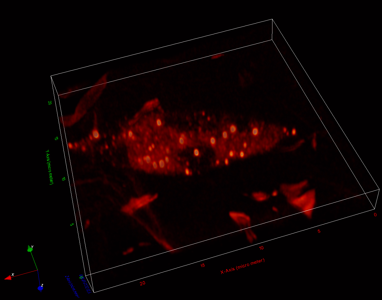
(Obj1) we will quantify phyB PBs features (size, intensity) and positioning relative to target loci and other nuclear compartments (heterochromatin and periphery) in intact leaf nuclei, comparing different light (Red versus Far-Red) and temperature (21℃ versus 30℃) conditions, using PHYB immunostaining, SABER-FISH labelling of PHYB target loci (S1), 3D STED imaging and available image analysis workflows2 (S2). (Obj2) We will measure the spatial distribution and temporal dynamics of phyB PBs relative to nucleosomes using live imaging (phyB and H2B fluorescent lines) and MINFLux super-resolution microscopy at the 10-50 nm scale (using SNAP-tagged phyB and immunostained H2B). To control the light and temperature conditions at the microscope we use a custom LED setup and a temperature control device (S3). Image analysis will be conducted using ChromTrack3D tools.
More information
Training benefits
Learning and applying super resolution imaging techniques (STED, MinFlux), in combination with high resolution FISH (SABER-FISH), to investigate whether phase separated phyB photobodies are directly associated with light-responsive chromatin loci.
You will also learn plant photobiology, live imaging of phyB photobodies, and principles of protein phase separation.
Requirements
Significant experience with confocal microscopy, and (plant) molecular biology. A Master in the field of cell or molecular biology. A desire to succeed in science.
Environment
Heidelberg (Germany) is one of the biggest molecular biology hubs in Europe. The Light Signaling and Cell Biology lab of Dr. van Gelderen is a young and ambitious lab with a friendly working environment and many contacts and collaborations within the institute, the Centre for Organismal Studies, and with the rest of Heidelberg University and the EMBL.
Responsible PI